| T O P I C R E V I E W |
| klinikaxp |
Posted - Oct 13 2020 : 17:56:57
I display dicom image. It requires modifications: sometimes user need to analyse bone structure or another part of body.
Do you have any experience in enhancing DICOM images?
I have spent many hours trying to use many functions and combinations of them, but the results are not satisfactory to the end user:
- the best effect gives a combination of Gamma Correction, Brightness and Contrast.
- sometimes Autoenhance3 allows to see more details.
- using Sharpen method degrades the quality
I tried to use Autoenhance1, Autoenhance2 and some other methods, but it is still not clear enough.
Maybe I should use histograms to focus on some ranges of brightness?
Maybe someone managed to achieve better results? Any tips?
I attach the sample of final image I would like to achieve: the bone structure has many details (some kind of net)
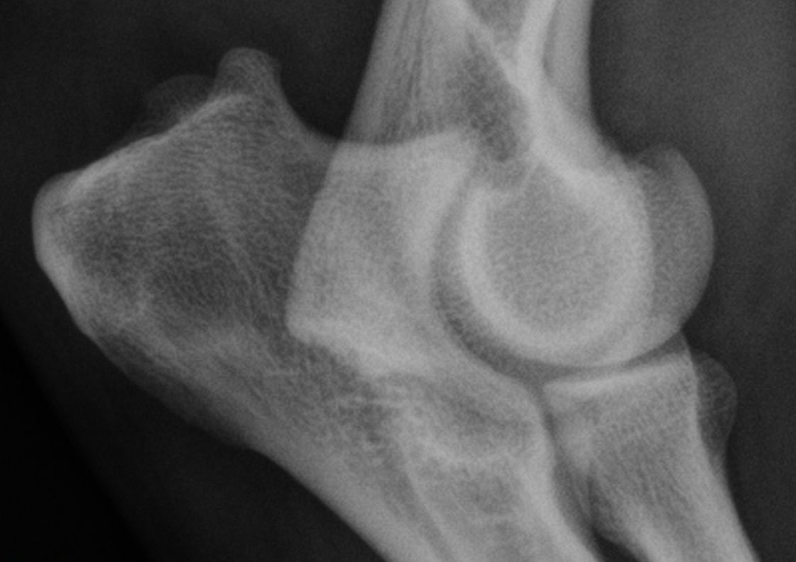 |
| 3 L A T E S T R E P L I E S (Newest First) |
| xequte |
Posted - Oct 13 2020 : 21:11:24
Hi
I don't think there is a perfect solution here, because we are dealing with gray levels, where the same gray can be found in both the subject (bone) and non-subject area.
Try using the TImageEnProc.DoPreviews and see what gives you the best result.
In this demo it is Image > Adjust Colors on the menu:
https://www.imageen.com/files/demos/run/ImageEditing/CompleteEditor/PhotoEn.exe
Nigel
Xequte Software
www.imageen.com
|
| klinikaxp |
Posted - Oct 13 2020 : 19:29:44
Nigel,
Thank you for quick response. I tried to modify channels in this way. But I still can not achieve the image with clear view. Please look at source dicom file (attached). Is it possible to modify this image to extract bone structure like in previous image?
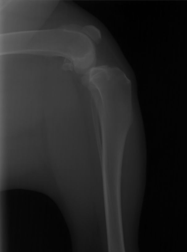 |
| xequte |
Posted - Oct 13 2020 : 18:41:50
Hi
How about adjustment of levels?
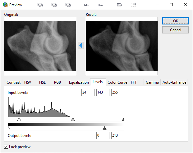
Nigel
Xequte Software
www.imageen.com
|